A laboratory model made from human lung tissue has preserved more than lung cells. It has preserved part of the lung’s local immune system—and allowed researchers to watch that system respond to SARS-CoV-2.
The study, published October 7 in Nature, describes long-term three-dimensional cultures made from intact adult distal-lung tissue. The organoids retained epithelial and stromal components along with T cells, B cells, natural killer cells and myeloid cells. When infected with SARS-CoV-2, they produced inflammatory signals and virus-specific T-cell responses. When researchers stimulated the organoids with SARS-CoV-2 peptide pools before a later infection, the subsequent infection was reduced.
That is a meaningful advance in experimental biology. It is also easy to overstate.
The study does not show that scientists have created a vaccine, that an inhaled vaccine based on the method will work in people, or that lung-resident T cells can protect humans independently of antibodies and circulating immune cells. Its strongest result is narrower and more useful: researchers now have a more integrated laboratory window into how human lung tissue and resident immune cells respond to respiratory infection.
The study is described in the published Nature paper, and the National Institutes of Health has summarized the work as an NIH-supported effort using lung fragments from more than 220 patient volunteers. The distinction between what the model demonstrates and what it may eventually enable is central to understanding the result.
Why tissue-resident immunity is difficult to study
Respiratory infections begin at surfaces where the body meets the outside world. Yet much immunological research relies on blood samples, isolated cells or simplified cultures. Those systems are useful, but each leaves something out.
A blood sample can show circulating antibodies and immune cells. It cannot fully reproduce what happens inside lung tissue. An epithelial culture can show how respiratory cells become infected. It may not reveal how resident immune cells respond in the same local environment. Animal models can capture whole-body interactions, but they do not always reproduce human immune biology.
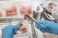
The Stanford-led study attempts to preserve several of those layers together. The researchers began with intact fragments of adult human distal lung and maintained them in a three-dimensional air–liquid interface culture. The resulting organoids retained multiple cell types from the original tissue rather than adding an immune component as a separate afterthought.
That design matters because tissue-resident immune cells are defined partly by their location. They are positioned in tissues where pathogens may first appear, and they can respond without waiting for an entirely new immune response to develop elsewhere. But lung-resident memory T cells remain difficult to study directly in living people, especially when researchers want to distinguish local activity from the contribution of circulating immune cells and secondary lymphoid organs.
What the experiment showed
The model preserved T cells with markers associated with tissue residence and memory, along with elements of the T-cell receptor repertoire found in the original lung tissue. It also maintained other immune populations, including B cells, natural killer cells and myeloid cells.
When the organoids were infected with SARS-CoV-2, the lung epithelium became infected and the tissue produced inflammatory signals. The researchers detected antigen-specific T-cell activity rather than only a generalized response to cellular damage. The model therefore provided evidence that resident immune cells could recognize viral material within the preserved lung environment.
The most consequential experiment came next. The researchers exposed intact organoids to pools of SARS-CoV-2 peptides, then challenged them with the virus. In the model, that stimulation activated resident T cells and reduced subsequent viral infection.
This supports the possibility that local immune memory can alter the response of lung tissue without the presence of a complete secondary lymphoid system. It also offers researchers a way to study tissue-resident immunity in a setting that is more integrated than a single-cell assay.
But “reduced infection in an organoid” is not the same result as “prevented disease in a person.” The measured outcome is important precisely because it is limited and defined.
A peptide experiment is not a vaccine trial
The phrase “local vaccination” can make the study sound closer to clinical application than it is. The researchers did not administer an approved vaccine to a person. They stimulated organoids with SARS-CoV-2 peptide pools under controlled laboratory conditions.
That intervention bypasses many of the practical problems that a vaccine must solve. A real vaccine must reach the intended tissue, stimulate the appropriate cells, produce an immune response of useful strength and duration, avoid excessive inflammation and work within the complicated environment of a living body. It must also be tested for safety, dose, delivery and effectiveness across diverse people.
The organoid experiment does not answer those questions. It shows that local antigen stimulation can activate resident T cells under the conditions established in the model. It does not establish that an inhaled or intranasal vaccine can reproduce the result in people, nor that such a vaccine would be better than existing approaches.
Nor does the study show that T cells acted alone. The model includes several immune populations, and the experiments do not turn lung-resident T cells into a complete explanation for protection against respiratory disease. In living people, local immunity operates alongside antibodies, circulating immune cells, airway defenses and systemic responses.
Human-derived is not the same as human
The model’s use of human tissue improves its relevance. It does not remove the limitations of an artificial system.
The organoids do not reproduce an intact body’s blood flow, lymphatic drainage, breathing mechanics, nervous-system signaling, endocrine environment or communication with distant immune organs. They cannot reproduce the complete sequence of exposure, symptoms, recovery and long-term disease. Their biological realism is therefore selective rather than total.
Donor variation is another important issue. The tissue came from people with different ages, medical histories, prior infections and vaccination backgrounds. That variation may eventually make the system valuable for comparing immune responses among individuals. It also complicates generalization. A striking response in one donor-derived line may not represent the average response across people.
The viral experiments were conducted with SARS-CoV-2. That makes the system relevant to coronavirus research, but it does not establish that the same response will occur with influenza, respiratory syncytial virus, bacterial infections or future pathogens. Each organism interacts with lung tissue differently, and each may require separate validation.
The relevant question is not whether the organoid looks sophisticated. It is whether the model reliably answers a defined scientific question.
A concrete test of the organoid promise
That question connects this study to the broader institutional shift toward human-based biomedical research. In a September 30 WBC article, I examined the growing investment in organoids, tissue chips and other new approach methodologies. The central issue there was validation: whether a particular method produces reliable information for a particular scientific or regulatory decision.
The lung study provides a useful case study because it demonstrates both the promise and the difficulty of that standard.
The model may help researchers investigate how respiratory viruses interact with lung tissue, compare local immune responses among donors, study chronic lung disease and test candidate interventions before moving to animal or clinical research. It may also help identify questions for which animal models are poorly matched to human tissue-resident immunity.
But a credible validation program would need more than another striking image or another successful infection experiment. Researchers will need to replicate the findings across laboratories, standardize culture conditions, compare the organoids with fresh human tissue and determine how well model results correspond to clinical observations.
They will also need to report where the model fails. A platform is more useful when researchers know the boundaries of its predictive power. That requires studying inconsistent results, donor lines that respond differently and interventions that appear promising in the dish but do not translate to living systems.
The commercial context matters, but it does not decide the science
The paper discloses intellectual-property interests involving the methods, along with commercial or advisory relationships for some authors. Those disclosures do not invalidate the findings. They do, however, make careful language about future applications especially important.
A patent is not evidence that a medical product will work. A promising model is not evidence that a vaccine platform is ready for human testing. Translational claims should be evaluated through independent replication, comparative studies and clinical evidence rather than through the existence of a plausible mechanism or commercial interest.
This is not an argument against developing the technology. It is an argument for distinguishing research capability from medical performance.
The immediate achievement is epistemic
The most defensible conclusion is that scientists now have a more realistic laboratory system for studying part of the lung’s immune life. Human lung tissue maintained outside the body can preserve resident immune cells, respond specifically to SARS-CoV-2 and reduce later viral infection after local antigen stimulation.
That result does not produce a vaccine. It does not replace clinical research. It does not show that tissue-resident T cells are the dominant source of protection against respiratory disease.
It does something less dramatic and more valuable: it makes a previously difficult biological question easier to investigate. Researchers can now ask how local immune memory behaves within preserved human lung tissue, how much responses vary between donors and whether the same mechanisms appear with other pathogens or interventions.
The next question is not whether the organoid is a human lung. It plainly is not. The question is whether it reproduces the particular lung functions scientists need to study with enough reliability to improve biomedical decisions.
That answer will come from replication, comparison and clinical relevance—not from the novelty of the model itself.